


The health of one’s eyes is crucial to one’s overall well-being because they serve as the window to the world. The fundus examination is one of the most potent medical tools used in the contemporary practice of eye exams. The special blood test enables the physician to examine carefully the back of your eye (also called the fundus) that contains the retina, macula, optical disk, and blood vessels of the retina.
When utilizing advanced fundus-examining technologies like digital retinal imaging and ophthalmoscopy, fundus examinations can detect the onset of possible vision-threatening diseases like glaucoma, diabetic retinopathy, and macular degeneration early enough to potentially avert the symptoms, long before these may be evident. It is also useful in the diagnosis of changes occasioned by seasonal diseases like hypertension and diabetes.
Our vision at Dr. Aggarwal’s Eye Clinic is to save and safeguard your eyes by detecting problems and arriving at a proper diagnosis at an early stage. Our state-of-the-art equipment and highly skilled ophthalmologists ensure accuracy, comfort, and effectiveness when conducting retinal evaluations on patients.
What is Fundus Examination?
A fundus Examination is A special diagnostic eye examination that enables an eye specialist (ophthalmologist) to look at the inner (or back) surface of the eye (also known as the fundus). The evaluation is carried out to examine the status of the retina, macula, optic disc, and retinal blood vessels, where healthy vision requires clear and healthy individuals.
- Retina: The light-sensitive tissue at the back of the eye that uses light to create images through visual signals and makes us see. If retinal damage is not addressed, it may worsen and result in permanent blindness instead.
- Macula: A very small but important part of the retina involved in short, detailed sight, utilized in reading, driving, and recognizing faces.
- As the optic nerve leaves the eye, the visual information is transformed and transmitted to the brain via the optic disc. The development of alterations in the optic disc could increase glaucoma or other optic nerve pathologies.
- Retinal Blood Vessels: The system of small arteries and veins that deliver oxygen and other nutrients to the retina; an abnormal appearance here would indicate the presence of diabetic retinopathy, hypertensive retinopathy, or vascular occlusion.
Ophthalmologists may also identify preliminary stages of retinal disorders, macular degeneration, and optic nerve disorders with the help of such instruments as direct ophthalmoscopy, indirect ophthalmoscopy, and digital retinal imaging. The test is not invasive, simple, and constitutes a very crucial component of the complete eye examinations, particularly for individuals who have diabetes, high blood pressure, or who have relatives with eye disorders.
What is Fundus Examination Important?
Most of eye diseases develop silently and run without apparent signs until poor eyesight impairs vision strongly. Such conditions can be identified at the early stages with a regular fundus check.
The most important advantages are:
- Early Diagnosis of Eye Diseases: Eye conditions like glaucoma, retinal detachment, and diabetic retinopathy.
- Vision loss prevention occurs due to early detection, leading to early treatment and thereby the prevention of vision loss.
- Diagnosis of Body Disorders: Information about some of the conditions, such as high blood pressure and diabetes, can be observed in the retina.
- Disease Progression: It can be of assistance in monitoring the progression of diseases in patients who have chronic eye diseases.
Fundus Examination Types
1. Direct Ophthalmoscopy
- It is done with a handheld ophthalmoscope.
- Gives an enlarged picture of the central retina.
- Appropriate when fast, genuine inspections are needed, such as in clinics.
2. Indirect Ophthalmoscopy
- Carried out using a device mounted on the head and a condensing lens.
- Increases the field of vision and enables investigation of the peripheral retina.
- In particular, it is used to visualize retinal tears and detachment.
3. Fundus Photography
- Takes high-resolution images of the retina with the use of digital retinal imaging.
- Provides the possibility to accurately document and track over time.
- Beneficial to the patient education as well as long-term follow-up.
Procedure of Fundus Examination, Step-by-Step
An essential diagnostic method for evaluating the health of the macula, blood vessels, optic disc, and retina is a fundus examination. It is significant in identifying early symptoms of diseases like diabetic retinopathy, glaucoma, macular degeneration, and retinal detachment. Now, let us take a closer look at how the process will take place in a step-by-step process:
1. Patient Preparation
A properly illuminated examination room will have you seated in relative comfort. Your medical and eye records, as well as your symptoms such as blurred vision, floaters, and eye pain, will be checked by the ophthalmologist. This will assist in designing the examination according to your requirements.
2. Pupil Dilation
Using certain dilating eye drops is necessary to enlarge the pupils. This gives the doctor a panoramic view of the structures of the retina. The drops can result in moments of light sensitivity or blurred near vision, but usually subsides after a few hours.
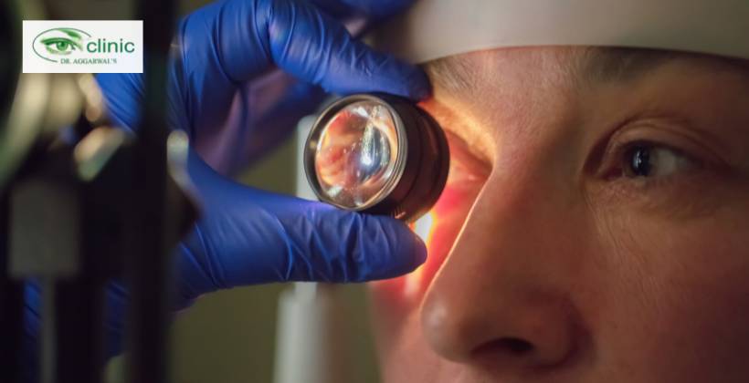
3. Detailed Examination
With an ophthalmoscope (and/or an advanced fundus camera), the ophthalmologist examines the retina, macula, optic nerve head, and retinal blood vessels. This is done to identify abnormalities that may include bleeding, swelling, and degeneration.
4. Image Documentation
Stereoscopic photographs at high resolution can be taken of the retina. These are saved and compared in the future so that the doctor can keep track of any development of the disease over time.
5. Report & Recommendations
The outcome of the examination is explained by the ophthalmologist after the examination. Should any disease of the retina be identified, an individualized treatment regimen or schedule will be recommended.
Conditions that are identified as a result of the Fundus Examination
This examination may show:
- Diabetic Retinopathy: Diabetes damages the retina’s blood vessels.
- Glaucoma: The pressure in the eye is normally elevated, triggering damage to the optic nerve.
- Retinal Detachment: An urgent health care issue in which the retina comes off its supportive structure.
- Macular Degeneration: Progressive destruction of the macula, which is used to see central vision.
- Optic Neuritis: The inflammation of the optic nerve, which in some cases is associated with neurological diseases.
- Hypertensive Retinopathy: High blood pressure damage to the retina.
Advantages of periodic Fundus examinations
Regular eye exams aid in the early detection of eye problems and are crucial for maintaining eyesight. This easy but effective test enables ophthalmologists to evaluate the retina, macula, optic disc, and blood vessels to detect any levels of diabetic retinopathy, glaucoma, and the development of macular degeneration before any damage.
Key Benefits
- Prevention of Blindness: Early detection will save eyes and result in early treatment.
- Monitoring Eye Health: Monitors the changes in diabetic, hypertensive patients, and chronic diseases.
- Non-Invasive, Painless: It is a well-tolerated procedure that can be applied to all age groups.
- Fast and Precise Results: Fast Results to diagnose eye health and access treatment.
- Economic Medication: Eliminates the cost implications of going to treatment at a late stage.
Through the incorporation of regular fundus examinations, all individuals can use routine eye check-ups to protect their vision, keep their eyes healthy, and have a better life.
Reasons to do a Fundus Examination in Dr. Aggarwal Eye Clinic
In Dr. Aggarwal Eye Clinics, we use experience, exactness, as well as technology to do precise fundus examinations.
We have the following advantages:
- Cutting-edge Diagnostic instruments: The Newest fundus camera and ophthalmoscopes.
- Experienced Ophthalmologists: Qualified doctors who have many years of clinical practice.
- Patient-Centered Care: Open communication and a less stressful testing environment.
- Low Price: Reasonable charges for high-quality care.
We strive to make sure that all our patients are in full possession of their eye health and understand that they are receiving optimal care.
FAQs
Q1: At what interval ought I to have a fundus?
Ans: Normal adults are advised to have one annually, whereas individuals with diabetes, high blood pressure, or any eye condition may be required to have them more frequently.
Q2: Does the procedure hurt?
Ans: No, it is entirely non-invasive and painless.
Q3: Does it have the ability to detect diabetic eye damage?
Ans: Yes, it is one of the finest methods of responding to diabetic retinopathy at an early point.
Q4: How long does the dilation effect last?
Ans: On average, 4-6 hours, when the near sight can be blurred and sensitivity to light can increase.
Q5: Does fundus photography apply to everybody?
Ans: These are not required as they are advised to be documented and compared when one visits again.
Conclusion
A fundus examination is not a regular eye check: it is a look at your overall health. Identifying symptoms of vision-threatening disease and systemic diseases early will enable you to deal with the condition before irreparable deterioration has taken place.
Using the most modern equipment, highly-skilled physicians, and a patient-centered attitude, Dr. Aggarwal Eye Clinic makes sure that each retinal examination is precise, thorough, and painless. It is not necessary to wait till symptoms manifest, but the fundus examination should be conducted today to preserve your vision in the future.